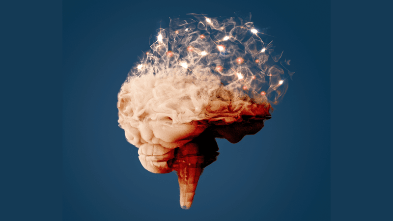

Bioengineers have created a soft, thin, and stretchable bioelectronic device that can be implanted into frog embryos’ developing brains, effectively making tiny cyborgs. This technology, they suggest, could help us understand and treat neurodevelopmental conditions in children in the future.
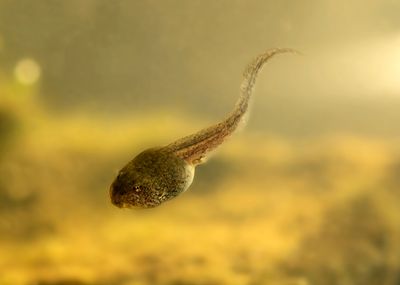
The device is implanted into the embryo's neural plate, a flat structure that eventually folds into what is known as the neural tube, two precursors in the development of the brain and spinal cord in vertebrates. Although it sounds dangerous, the researchers have shown that the device can be easily integrated into the animal’s brain as it develops, allowing them to continuously monitor brain activity during the embryo’s subsequent development.

Before this latest investigation, if researchers wanted to record the electrical activity of single neurons with high resolution, they would have to implant patch-clamps or metal electrodes into mature brains. As you can imagine, this is a pretty invasive process and not one that is free of risks.
The team behind the new probe had previously created tissue-like microelectronics that have made single-cell brain recording less invasive. But even these developments are not perfect; neurons connect with each other at nanometer resolutions (basically scales that are a billionth of a meter), so no matter how soft or gentle a probe is, it will still cause some damage. The new method helps to minimize this.
“If we can fully leverage the natural development process, we will have the ability to implant a lot of sensors across the 3D brain noninvasively, and at the same time, monitor how brain activity gradually evolves over time,” said study author Jia Liu in a statement. “No one has ever done this before.”
Reaching this point has taken years of effort by Liu and colleagues, but now they have a soft, flexible, non-invasive bioelectronic probe for the developing brain, which, they say, has the consistency of tofu (yum).
Prior to this, the team had embedded electrode arrays into petri dishes of stem cells that stretched and folded with the tissues as they grew. They basically became cyborg heart and brain organoids. However, it turns out there are important differences between organoids and amphibian embryos that pose significant challenges.
“It turns out tadpole embryos are much softer than human stem cell-derived tissue,” he explained. “We ultimately had to change everything, including developing new electronic materials.”
To overcome this, the team produced new implants using fluorinated elastomers – synthetic polymers – which are as soft as biological tissues but are highly resilient and can withstand nanofabrication processes. They also contain multiple sensors for recording brain activity, despite their tiny size.
During a conversation with IEEE Spectrum, Liu emphasized that the development of these probes is not intended for use on human embryos. That, the researcher said, would be “completely unethical and not our intention at all”. Still, the team hopes this work will help inform a better understanding and potentially treatment of neurodevelopmental conditions.
“Autism, bipolar disorder, schizophrenia – these all could happen at early developmental stages,” explained Liu in the previously mentioned statement. “There is just no ability currently to measure neural activity during early neural development. Our technology will really enable an uncharted area.”
The study is published in Nature.